At Safe Heart, Dr. Amit Gupta, an experienced Heart Valve Specialist in Jaipur, provides
expert evaluation and personalized guidance for people with heart valve conditions. He takes time to
understand each patient’s concerns, explains suitable options clearly, and helps plan care based on the
valve problem, overall health, and individual needs.
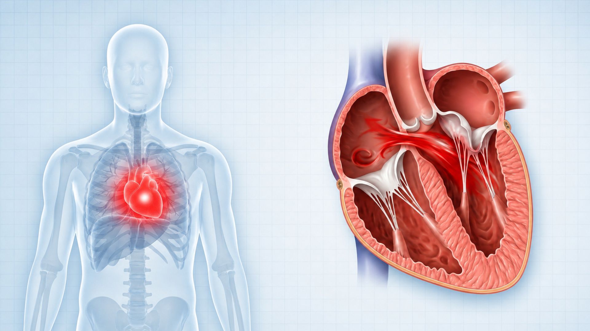
Heart valve problems can change the way blood flows through the heart and may lead to
breathlessness, chest discomfort, tiredness, dizziness, or swelling. As a Heart Valve Doctor in Jaipur, Dr.
Amit Gupta focuses on understanding the cause and severity of each valve problem before discussing the most
suitable care for the patient.
AorticSevere aortic
stenosis
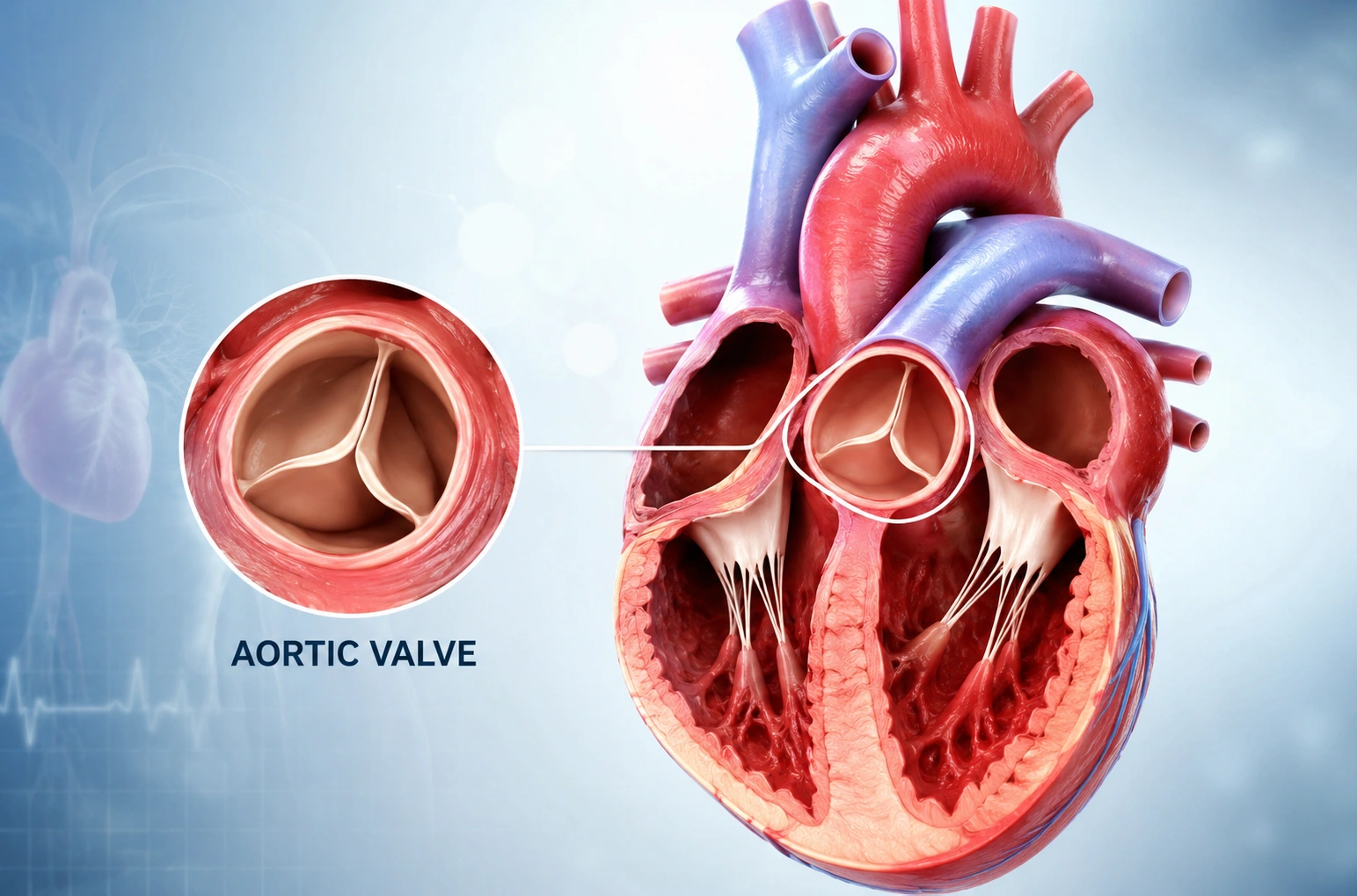
The aortic valve becomes narrow and does not allow blood to flow out of the heart easily. It can cause
breathlessness, chest pain, dizziness, or fainting, especially when the narrowing becomes severe.
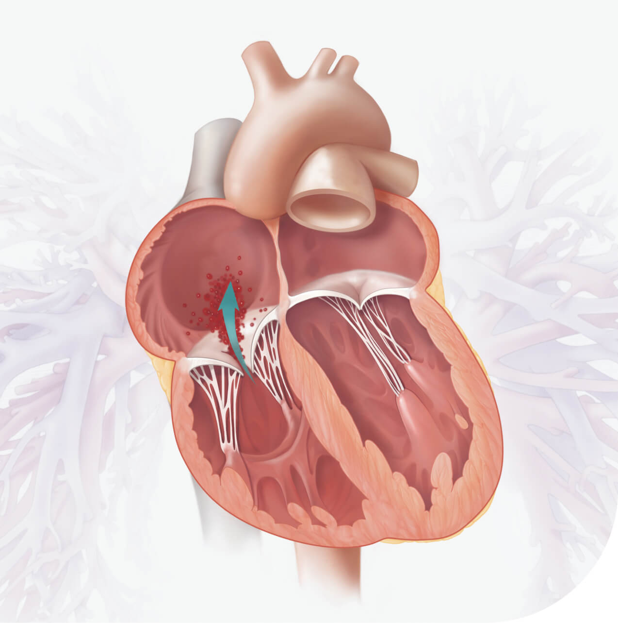
AorticAortic regurgitation
The aortic valve does not close properly, so some blood flows backward into the heart. Over time, this
can put extra pressure on the heart and may affect how well it pumps.
MitralMitral regurgitation
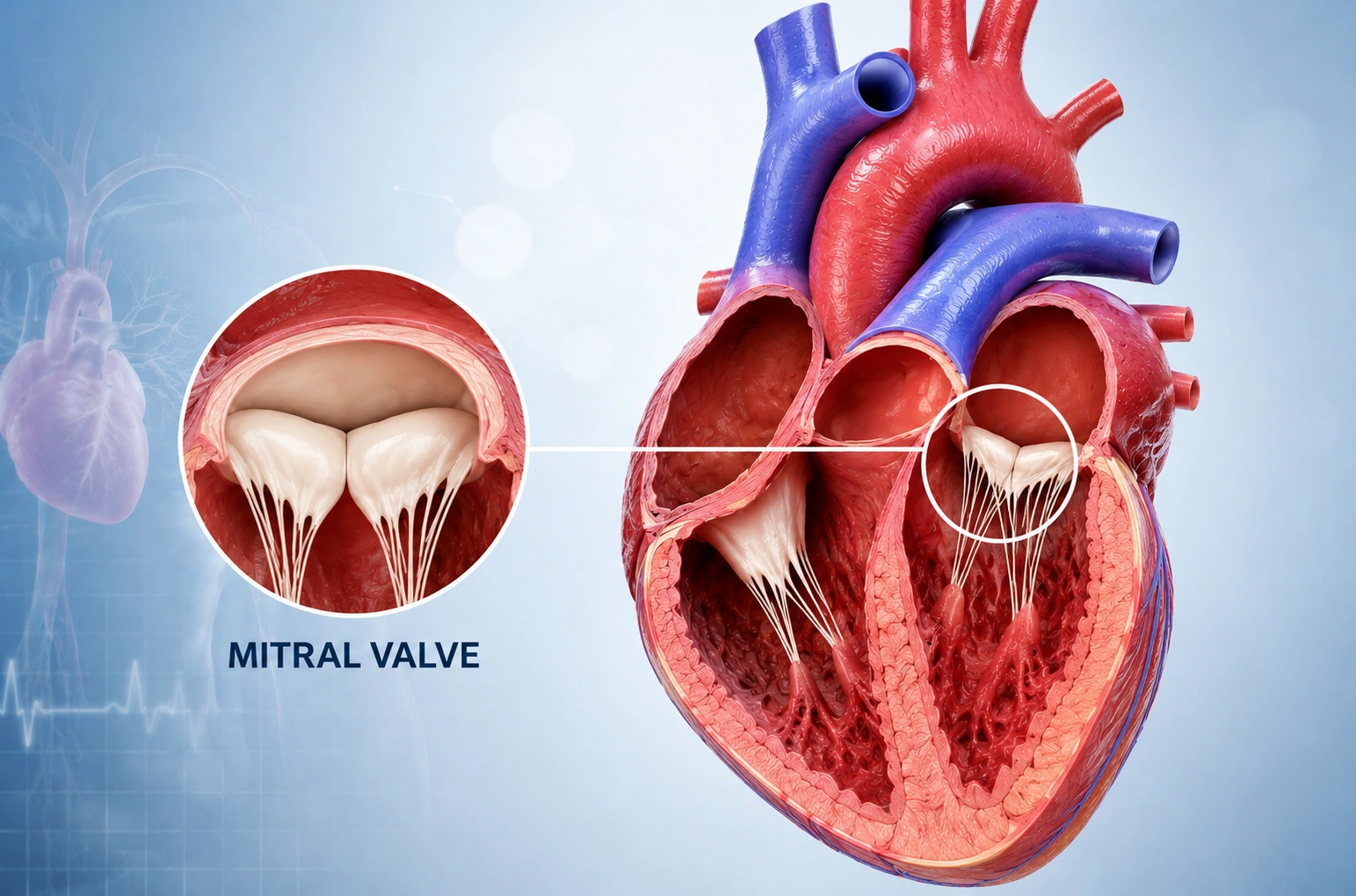
The mitral valve becomes leaky, allowing blood to flow backward instead of moving forward normally. Some
people have no symptoms, while others may develop breathlessness, tiredness, or reduced ability to do
everyday activities.
MitralRheumatic mitral stenosis
This condition can develop after rheumatic heart disease causes the mitral valve to become narrow. The
narrowed valve makes it harder for blood to pass through the heart and may cause breathlessness,
tiredness, or a fast heartbeat.
TricuspidTricuspid regurgitation
The tricuspid valve does not close properly, allowing blood to flow backward. When the leakage is
significant, it may cause swelling in the legs or abdomen, tiredness, breathlessness, or other symptoms.
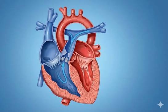
PulmonaryPulmonary stenosis
The pulmonary valve becomes narrow, making it harder for blood to travel from the heart to the lungs. It
is often present from birth and, when significant, may need treatment such as balloon valvuloplasty.
Four valves, four failures
Which Valve Is Affected?
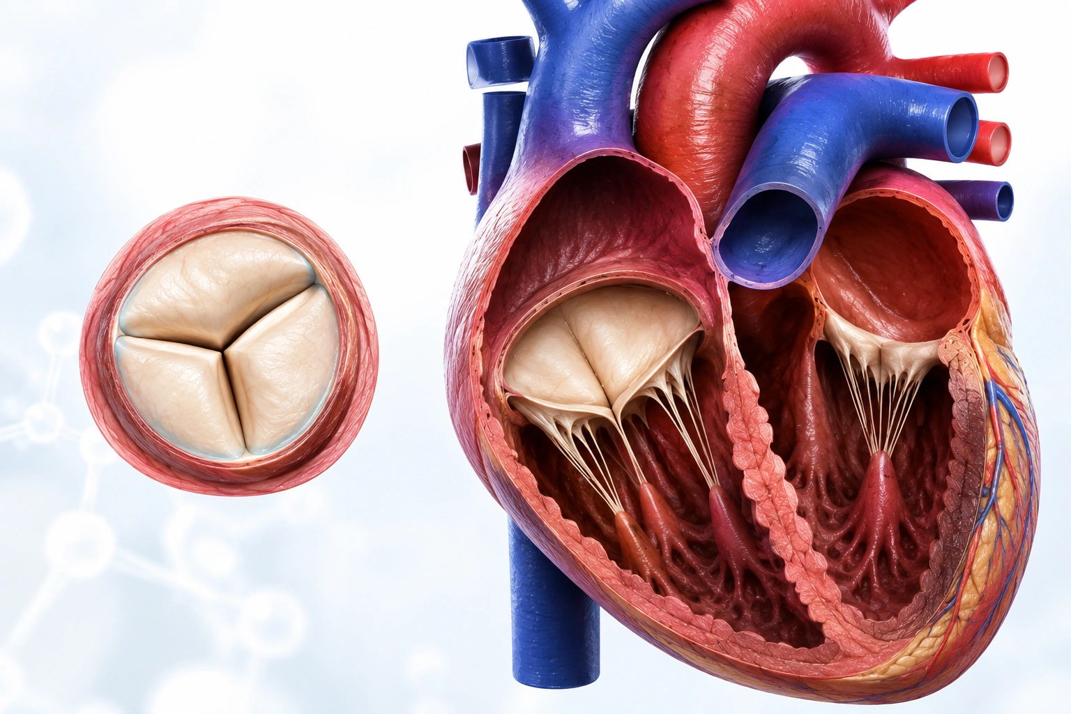
The heart has four valves that keep blood moving in the right direction. When a valve becomes
narrow or does not close properly, blood flow can be affected, and symptoms may develop. At Safe Heart, Heart
Valve Treatment in Jaipur starts with understanding which valve is affected, how severe the problem is, and
what care may be suitable.
Aortic valveNarrowing treated by
TAVI
1
Aortic Valve
Most common after age 65
The aortic valve allows blood to leave the heart and travel to the rest of the body. Aortic stenosis
makes the valve narrow, often because of calcium buildup, while aortic regurgitation causes blood to leak
backward when the valve does not close properly.
SignsBreathlessness, chest discomfort, dizziness, or fainting
TreatmentDepending on the severity and the patient's overall health,
treatment may include medicines, TAVI, or surgical valve replacement.
2
Mitral Valve
Leaks in the elderly, rheumatic in the young
The mitral valve helps blood move from the upper to the lower chamber on the left side of the heart.
Mitral stenosis makes the valve narrow, while mitral regurgitation causes blood to leak backward.
Rheumatic heart disease is an important cause of mitral stenosis.
SignsBreathlessness, tiredness, palpitations, or difficulty with physical
activity
TreatmentDepending on the cause and severity, options may include medicines,
valve repair or replacement, MitraClip/TEER for suitable patients, or balloon mitral valvuloplasty for
selected cases of rheumatic mitral stenosis.
3
Tricuspid Valve
The one most often overlooked
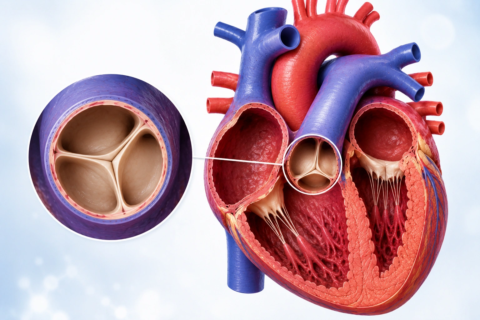
The tricuspid valve sits on the right side of the heart and helps blood move toward the lungs. Tricuspid
regurgitation occurs when the valve does not close properly, allowing blood to flow backward. Significant
leakage can lead to fluid buildup and strain on the right side of the heart.
SignsSwelling in the legs or abdomen, tiredness, or breathlessness
TreatmentA Heart Valve Specialist in Jaipur may recommend medicines, surgery,
or a suitable catheter-based procedure depending on the cause, severity, symptoms, and overall
health.
4
Pulmonary Valve
Usually present from birth
The pulmonary valve controls blood flow from the right side of the heart toward the lungs. Pulmonary
stenosis makes the valve opening narrower and is often present from birth, although some people may be
diagnosed later.
SignsBreathlessness during activity, tiredness, dizziness, or a heart
murmur
TreatmentBalloon valvuloplasty may be considered for suitable patients when
pulmonary valve narrowing is significant. The appropriate approach depends on the valve condition and
its severity.
Which Treatments Are Available for Heart Valve Problems?
For patients considering heart valve surgery in Jaipur, the right treatment depends on the valve
affected, how severe the problem is, symptoms, overall health, and valve anatomy. Depending on these factors,
care may involve medicines, valve repair, valve replacement, or a catheter-based procedure.
AorticTAVI /
TAVR
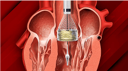
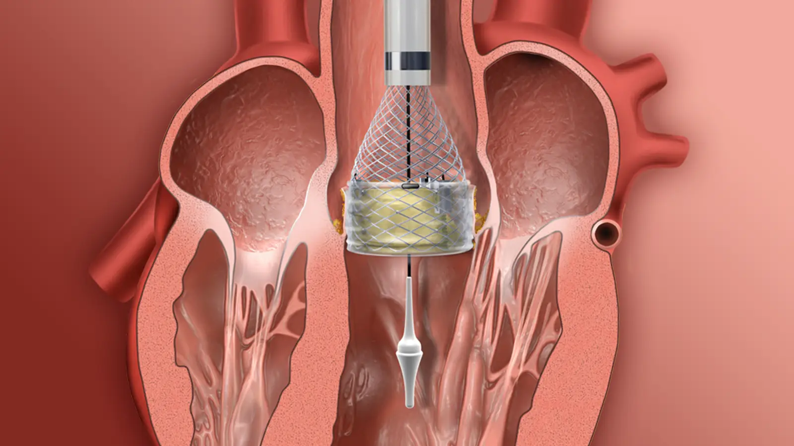
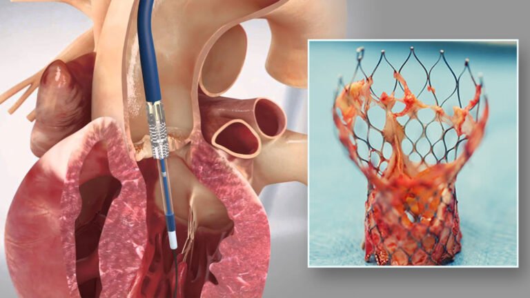
TAVI, also called TAVR, replaces a diseased aortic valve without opening the chest. A new valve is guided
through a blood vessel and placed inside the old valve. It may suit selected patients with severe aortic
stenosis.
MitraClip is a minimally invasive way to repair a leaking mitral valve. A small clip brings the valve
leaflets closer together, helping reduce blood leakage. It may be considered for selected patients with
significant mitral regurgitation.
It helps open a narrowed heart valve. A small balloon is guided through a catheter and gently inflated at
the narrowed area. It is commonly used for suitable rheumatic mitral stenosis and selected pulmonary
stenosis.
They replace the mitral or tricuspid valve using a catheter rather than conventional open surgery. These
are advanced options for selected patients when the valve problem and overall health make the procedure
appropriate.
When a previously implanted biological valve starts to fail, a new valve can sometimes be placed inside
it through a catheter. This valve-in-valve approach may help selected patients avoid another major valve
operation.
Catheter procedures are not right for everyone. In some patients, surgery may offer the better option.
Depending on the valve problem, the surgeon may repair the existing valve or replace it with a new
mechanical or biological valve.
Need a Second Opinion for Your Heart Valve Problem?
Get a second opinion from Dr. Amit Gupta, Heart Valve Specialist in Jaipur, before deciding on surgery or
another valve procedure. Your reports can be reviewed to understand your condition and suitable treatment
options.
Dr. Amit Gupta is a Heart Valve Specialist in Jaipur with specialised training in structural heart
intervention. He completed his DM in Cardiology from G. B. Pant Hospital, New Delhi, and later completed a
Structural Heart Intervention fellowship at John Hunter Hospital, Australia.
His current expertise includes TAVI, transcatheter edge-to-edge repair (TEER), MitraClip, and transcatheter
mitral valve replacement (TMVR), along with other structural and interventional cardiac procedures.
Dr. Amit Gupta, a Heart Valve Specialist in Jaipur, has received international training in
structural heart care and advanced transcatheter valve procedures through recognised clinical and technology
programmes.
John Hunter Hospital
MitraClip™ G5 Implanter
Edwards SAPIEN 3 Ultra
Abbott Navitor Vision™
मरीजों के लिए
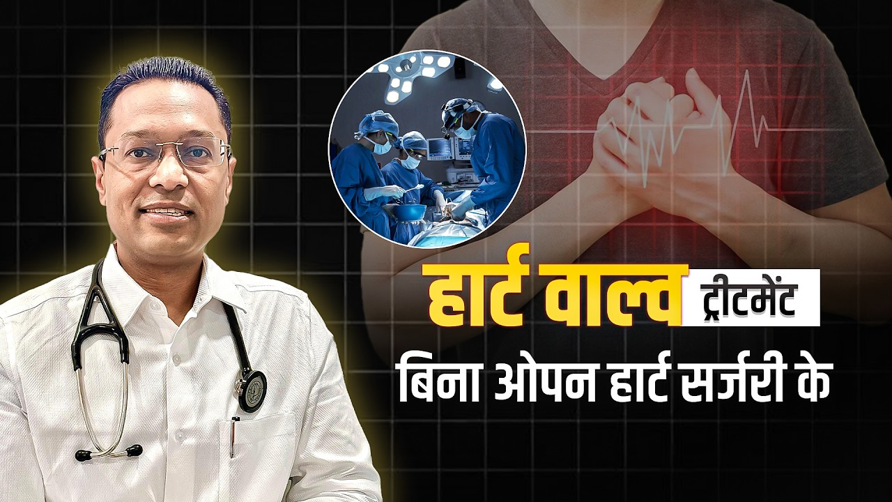
बिना ओपन सर्जरी वाल्व का इलाज
कुछ मरीजों में खराब या लीक हो रहे वाल्व का इलाज बिना छाती खोली भी किया जा सकता है। Heart valve replacement
in Jaipur के लिए TAVI जैसे कैथेटर-आधारित विकल्प उपयुक्त मरीजों में इस्तेमाल किए जा सकते हैं।
TAVI — खराब एओर्टिक वाल्व को कैथेटर से बदलने का विकल्प
MitraClip — लीक हो रहे मिट्रल वाल्व की लीकेज कम करने का विकल्प
कम इनवेसिव इलाज — कुछ प्रक्रियाओं में बड़ी सर्जरी की जरूरत नहीं होती
A Heart Valve Treatment in Jaipur plan depends on the valve problem, age, overall health, heart
anatomy, and surgical risk. A detailed evaluation helps decide whether TAVI or open surgery may be more
suitable.
Comparison of TAVI and open-heart aortic valve replacement
What you want to know
TAVI / TAVR
Open Surgery
Chest incision
No large chest opening; the valve is usually delivered through a blood vessel.
The chest is opened to reach and replace the diseased valve.
Anaesthesia
Sedation or general anaesthesia may be used, depending on the procedure.
General anaesthesia is normally required.
Recovery
Recovery is usually quicker, although it varies between patients.
Recovery usually takes longer because the chest needs time to heal.
Hospital stay
Usually a shorter hospital stay when recovery is uncomplicated.
Usually requires a longer hospital stay for surgical recovery.
Who may be suitable?
May suit selected patients based on age, health, anatomy, and surgical risk.
May suit patients when surgery offers a better option based on their condition and anatomy.
Valve durability
Good durability data are available, while very long-term evidence is still developing.
Long-term durability is well established, depending on the valve type used.
If the valve fails later
A valve-in-valve procedure may be possible for selected patients.
Further surgery or another treatment may be considered depending on the situation.
Still unsure which option may be right for you? Make an appointment with Dr. Amit Gupta,
Heart Valve Specialist in Jaipur, to discuss your reports and treatment options.
What happens next
Your Treatment Path
For Heart Valve Treatment in Jaipur, the journey usually starts with understanding your symptoms
and previous reports. The tests you need are then chosen to understand the valve clearly and plan suitable
care.
01
Consultation
Dr. Amit Gupta discusses your symptoms, medical history, previous reports, and examination findings to
understand your valve problem.
02
Echocardiogram
A 2D echo with Doppler shows how your heart and valves are working and helps assess how narrow or leaky a
valve has become.
03
Planning Scans
If more detail is needed, tests such as a CT scan or transesophageal echo (TEE/TOE) may be used to
understand the valve and plan a procedure.
04
Heart Team Review
For complex valve problems, cardiologists, surgeons, and imaging specialists review the findings together
and discuss which treatment may be most suitable.
05
Procedure & Follow-Up
Once the treatment is planned, the appropriate procedure is performed, and your recovery is monitored.
Follow-up visits and heart scans are arranged according to your condition and treatment.
60-second self-check
Do You Need a Valve Check?
Heart valve problems can develop slowly, and some people may have no symptoms at first. This
quick check can help you notice symptoms that are worth discussing with a doctor.
Not sure if your symptoms could be related to your heart valve?
Select anything that applies:
I get short of breath more easily than I used to
I feel chest pain, pressure, or tightness
I feel dizzy, lightheaded, or have nearly fainted
I have fainted or suddenly passed out
My heartbeat feels fast, fluttering, or irregular
I feel unusually tired or weak during normal activities
My feet, ankles, or abdomen become swollen
A doctor has told me that I have a heart murmur
I have had rheumatic fever or already have a heart valve problem
No symptoms selected yet
Select any symptoms or history that apply to you or the person you are looking for. This
check is for awareness, not diagnosis. If you have concerns, Dr. Amit Gupta, Heart Valve Specialist in
Jaipur, can assess your symptoms and advise whether further testing is needed.
A Heart Valve Doctor in Jaipur may recommend different tests depending on your symptoms, valve
condition, and the treatment being considered.
2D Echo
Shows your heart valves, their movement, blood flow, and how narrow or leaky a valve may be.
ECG
Checks your heart rhythm and looks for changes in your heart’s electrical activity.
TMT
Shows how your heart responds to exercise and can help assess symptoms that are difficult to notice at
rest.
Coronary Angiography
Checks the heart’s blood vessels for narrowing or blockages when coronary artery assessment is needed.
CT for TAVI
Provides detailed images of the aortic valve and blood vessels to help plan a TAVI procedure.
Holter & ABPM
Holter monitors your heart rhythm over time, while ABPM records your blood pressure during daily
activities.
Hear From Dr. Amit Gupta
Understanding Heart Valve Care
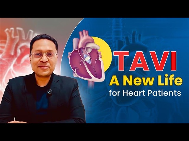
Dr. Amit Gupta, Heart Valve Specialist in Jaipur, explains heart valve problems, available
treatment options, and what patients should know before deciding on their care.
Patients sharing their experiences with Heart valve replacement in
Jaipur can help others understand what to expect from consultation, treatment, and follow-up care at Safe Heart.
★★★★★
Excellent serving Doctor. Honest, dedicated and very attentive towards their patient. For my 14-year-old
son, he acted as a god in saving his life. I recommend him as the best cardiologist in the city.
S
Sachin JakhetiaGoogle review
★★★★★
Dr Amit Gupta is one of the best cardiologists in Jaipur. He is such a humble person and has great
knowledge. It was a privilege to be treated by him as I am perfectly fine after his treatment.
R
Ram Prakash SharmaGoogle review
★★★★★
My mother has some heart-related problems. I visited many doctors but after meeting Doctor Amit my search
ended at Safe Heart. Very kind and helpful, immense knowledge. So thankful to Team Safe Heart.
K
Kapil SharmaGoogle review
★★★★★
Thanks for your good care and concern. Your manner put all patients at ease and helped them gain
confidence. You are truly a remarkable doctor and professional that we respect and trust.
A
Amar SharmaGoogle review
★★★★★
A very humble and honest cardiologist. For all my known patients I completely rely on him for best care.
God bless him and his family. Highly recommended for holistic cardiology care.
Meet Dr. Amit Gupta at Safe Heart Clinic for consultations and cardiac tests, while Manipal
Hospital provides advanced valve procedures and cath-lab care.
Safe Heart Clinic
Consultation · Echo · ECG
House No. 85, Sector 7, Vidyadhar Nagar, Jaipur 302039
Meet Dr. Amit Gupta, Heart Valve Specialist in Jaipur, for a detailed review of your
valve condition. Bringing your previous medical records can make the consultation more useful.
Previous heart reports and echocardiograms, especially your latest echo
CT, angiography, or other heart scans, if you have had them
ECG and other recent test reports related to your heart condition
A list of your current medicines, including their doses if possible
Details of previous heart procedures or surgeries, if any
A Heart Valve Specialist in Jaipur is a cardiologist with advanced experience in diagnosing and treating
heart valve problems. Dr. Amit Gupta evaluates narrowed and leaking valves and discusses options such as
TAVI, MitraClip, balloon valvuloplasty, valve repair, or replacement according to each patient's
condition.
What are the symptoms of heart valve disease?
Heart valve disease may develop gradually, so symptoms can be easy to overlook. Common symptoms include
breathlessness during activity, unusual tiredness, chest discomfort, dizziness, fainting, an irregular
heartbeat, and swelling in the feet, ankles, or abdomen. A heart murmur may also be detected during an
examination.
How is heart valve disease diagnosed?
A 2D echocardiogram with Doppler is the main test used to assess heart valves. It shows how the valves
move, how blood flows through them, and whether a valve is narrowed or leaking. ECG, CT, TEE, or other
tests may be added when needed.
Can a heart valve be replaced without open-heart surgery?
Yes, selected patients can have Heart valve replacement in Jaipur through catheter-based procedures such
as TAVI. A new valve is guided through a blood vessel and placed inside the diseased aortic valve.
Suitability depends on the valve problem, anatomy, health, age, and other factors.
What is the difference between TAVI and open-heart surgery?
Both can replace a diseased aortic valve, but they use different approaches. TAVI places the new valve
via a catheter, usually without opening the chest, whereas surgery involves reaching the heart through an
incision in the chest. The suitable option depends on your condition, anatomy, health, age, and surgical
risk.
Who may be suitable for TAVI?
TAVI may be considered for selected patients with significant aortic stenosis. The decision is based on
symptoms, echocardiogram and CT findings, valve anatomy, overall health, age, and surgical risk. A Heart
Team reviews these factors before deciding whether TAVI or another treatment is appropriate.
What is Valve-in-Valve treatment?
Valve-in-valve treatment places a new transcatheter valve inside a previously implanted biological valve
that has become narrowed or leaky. For selected patients, heart valve replacement in Jaipur may include
this less invasive option when the existing valve and anatomy are suitable.
What is MitraClip and when is it used?
MitraClip is a catheter-based procedure known as transcatheter edge-to-edge repair, or TEER. A small clip
helps the mitral valve leaflets close more effectively and reduces leakage. It may be considered for
selected patients with significant mitral regurgitation following a detailed valve assessment.
Is balloon mitral valvuloplasty still used in India?
Yes. Balloon mitral valvuloplasty remains an established treatment for selected patients with rheumatic
mitral stenosis. A balloon is guided through a catheter to the narrowed mitral valve and inflated to
improve the opening. Whether it is suitable depends on the valve structure and other clinical findings.
Does every heart valve problem need surgery?
No. Mild or moderate valve disease may only need regular check-ups, echocardiograms, and treatment of
related health problems. Heart valve surgery in Jaipur or a catheter-based procedure may be considered
when the disease becomes severe, symptoms develop, or the heart begins to show signs of strain.
What does heart valve treatment cost in Jaipur?
The Heart valve replacement cost in Jaipur varies according to the valve involved, procedure chosen, type
of valve or device, hospital stay, investigations, and individual needs. An accurate estimate can be given
after reviewing the patient's reports and deciding which treatment is appropriate.
What tests are needed before TAVI?
Before TAVI, an echocardiogram assesses the valve, while CT imaging shows the valve and blood vessels in
detail. ECG and other tests may also be advised. Heart Valve Treatment in Jaipur at Safe Heart Clinic is
planned after reviewing these findings and confirming whether TAVI is suitable.
Can I get a second opinion before heart valve surgery?
Yes. If you have been advised to undergo Heart valve operation in Jaipur, a second opinion can help you
understand the diagnosis and available options. Dr. Amit Gupta can review your reports and explain whether
surgery, TAVI, MitraClip, valvuloplasty, or another approach may be suitable.
How long does recovery take after a heart valve procedure?
Recovery varies depending on the procedure and the patient's overall health. Catheter-based procedures
generally involve less physical recovery than open surgery, but the exact time differs from person to
person. Your doctor will advise you about activity, medicines, work, driving, and follow-up after
treatment.
What should I bring to my heart valve consultation?
Bring your previous echocardiograms, ECG reports, CT or angiography reports, current medicines, and
details of any previous heart procedures or surgery. Having these records available helps your Heart Valve
Specialist in Jaipur understand your medical history and decide what further evaluation may be needed.
Why is early evaluation important for valve disease?
Heart valve disease can progress gradually, and some people may not notice symptoms initially. Regular
evaluation can help identify changes in valve function and heart performance. Seeing a Heart Valve
Specialist in Jaipur can help determine whether monitoring, medicines, or an intervention is appropriate.
Can heart valve disease be treated with medicines?
Medicines can help control symptoms and related conditions, but they usually cannot repair a severely
narrowed or leaking valve. Depending on the condition, heart valve treatment in Jaipur may involve
monitoring, medicines, valve repair, or replacement when an intervention becomes necessary.
When should I see a Heart Valve Doctor in Jaipur?
A Heart Valve Doctor in Jaipur can help if you have breathlessness, chest discomfort, dizziness,
fainting, palpitations, swelling, or a heart murmur. An evaluation can identify whether a valve problem is
present and whether you need further testing, regular monitoring, medicines, or a procedure.



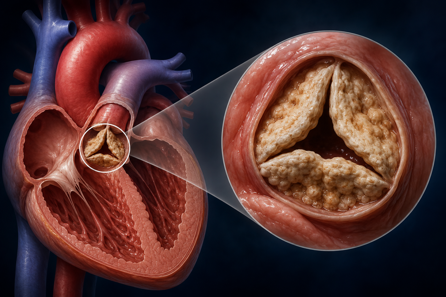 Severe aortic
stenosis
Severe aortic
stenosis